
HUMAN HEART :
Introduction : Human Heart
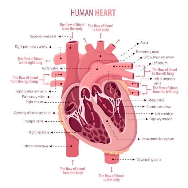
The human heart is a vital organ responsible for pumping blood throughout the body. It is a muscular organ located in the thoracic cavity, between the lungs, and slightly to the left of the center of the chest. The right atrium and right ventricle, as well as the left atrium and left ventricle, make up the four chambers of the heart. The atrioventricular groove, also known as the coronary sulcus, divides the atria from the ventricles on the surface, whereas interventricular grooves divide the ventricles from one another.
Shape and measurement of Human heart – Shape: conical or pyramidal.
Measurements: 12 cm is the length, width: 9 cm. Males weigh 300 g, while females weigh 250 g.
Diagram of Human Heart :
External features of Human heart :-
Human heart include external features:–
1. Apex
2. Base
3. Surfaces- 3 (Sternocostal, Diaphragmatic and Left surface )
4. Borders -4 (Right border, Left border, Upper border and inferior border )
1. Apex of the Human Heart :
The left ventricle forms a conical region at the top of the heart. Its direction is left, ahead, and downward. Situated 3.5 inches (9 cm) from the midline and slightly medial to the midclavicular line, it is situated at the level of the fifth left intercostal gap.
The point of maximum cardiac impulse (PMCI) or the outermost and lowermost push of the cardiac contraction (during ventricular systole) felt on the front of the chest is known as the apex beat. The apex beat is typically felt as a slight tap in the midclavicular line’s left fifth intercostal region.
Because the newborn heart is more horizontally positioned, its apex lies in the third or fourth left intercostal space. As a result, children up to the age of seven feel their apex beat in the third or fourth intercostal space, directly lateral to the midclavicular line.
2. Base of the Human Heart :
The base of the heart, sometimes referred to as the posterior surface, is composed of two atria, mostly the left atrium. Strictly speaking, the posterior surfaces of the left and right atriums make up two thirds and one third of the base, respectively. It faces opposite the apex and is pointed backward and to the right.
The following attributes of the base are distinctive:
1. It is located across from the apex.
2. In the prone position, it rests in front of the middle four thoracic vertebrae (T5–T8), and in the erect posture, it descends one vertebra (T6–T9).
3. Surfaces of the Human Heart :
1. Sternocostal (Anterior) Surface of Human Heart :- The right atrium and right ventricle, which are divided from one another by the anterior portion of the atrioventricular groove, make up the majority of its structure. The left ventricle and auricle both contribute to the formation of the sternocostal surface. The anterior interventricular groove divides the left and right ventricles.
2. Diaphragmatic (inferior) Surface of Human Heart :- This level surface lies atop the diaphragm’s major tendon. It is produced by the left and right ventricles working together, with the posterior interventricular groove serving as a barrier between them. Only the right ventricle makes up one-third of this surface, whereas the left ventricles make up the other two thirds.
3. Left surface of Human Heart :- The left ventricle forms the majority of it, with the left atrium and auricle contributing somewhat. It is pointed left, backward, and upward.
4. Borders of the Human Heart :
1. Right border of Human Heart : It is formed by the right atrium and is mostly vertical. It divides the base from the sternocostal surface and runs from the right side of the SVC opening to the IVC opening.
2. Left border of Human Heart :- It is bent and oblique. Its creation is aided by the left ventricle and left auricle. It divides the left and sternocostal surfaces as it goes from the left auricle to the heart’s apex.
3. Inferior border of Human Heart :- It runs from the IVC opening to the heart’s apex and is almost horizontal. The right ventricle forms it. This border also includes the right atrium. The diaphragmatic surface and the sternocostal surface are divided by the inferior boundary. It exhibits a notch known as the incisura apicis cordis close to the apex.
4. Upper border of Human Heart :- It is produced by the right and left atria, primarily the left one, and is slightly oblique. The pulmonary trunk and ascending aorta are in front of the top border, making it invisible from the sternocostal surface. A line connecting a point on the lower border of the second left costal cartilage, 1.5 inches from the median plane, and a point on the upper border of the third right costal cartilage, 1 inch from the median plane, can be used to identify it on the body’s surface.
Chambers of Human Heart :
Human heart consists four chamber
- Right Atrium
- Right ventricle
- Left Atrium
- Left Ventricle
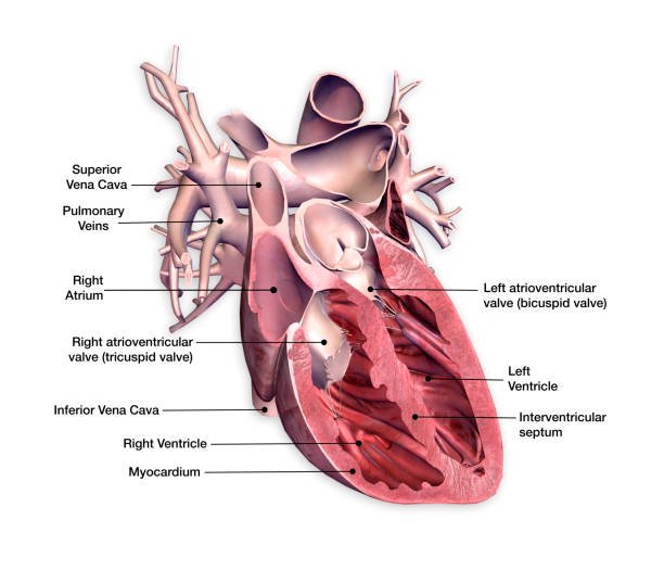
1. Right Atrium :
The right atrium, a thin-walled quadrilateral chamber located behind the right ventricle, plays a pivotal role in the circulatory system. Comprising a main cavity and a smaller outer portion known as the auricle, this chamber serves as the entry point for deoxygenated blood returning from the body.
External features of right atrium:-
1. The inferior vena cava (IVC) is located at the lower end of the extended right atrium, whereas the superior vena cava (SVC) is located at the top end.
2. The right auricle, or appendage, is formed by the extension of the top anterior section to the left. The auricle’s borders are notch-shaped. The infundibulum of the right ventricle and the roots of the ascending aorta are partially and totally overlapped by the right auricle.
3. Sulcus terminalis, a shallow vertical groove, runs along the right boundary between inferior and superior vena cavae. Located in the upper region of the sulcus is the sinuatrial (SA) node. It is equivalent to the crusta terminalis inside.
4. The right coronary artery and the tiny cardiac vein are lodged in the vertical right atrioventricular groove.
Internal features of right atrium:-
The right atrium’s interior is separated into two sections: the atrium proper (the rough anterior portion) and the sinus venarum (the main smooth posterior half). The two parcels are isolated by the crista terminalis. From inside, one may too see the right atrium’s septal divider. The right atrium’s septal wall develops from the septum primum and septum secundum. From inside the right atrium, the septal wall exhibits the following characteristics:
1. The fossa ovalis, a shallow depression in the lower section that resembles a saucer and is generated by the septum primum. It stands for the foetus’s foramen ovale location.
2. The distinct upper and lateral edge of the fossa ovalis is formed by the annulus ovalis/limbus fossa ovalis. It stands for the septum secundum’s free edge. The inferior border of the Annulus ovalis and the left end of the IVC valve are continuous.
3. The Triangle of Koch is a triangle region above the tendon of Todaro, a subendocardial ridge, beyond the anterior margin of the coronary sinus opening, and in front by the septal leaflet base of the tricuspid valve.
This triangle contains the atrioventricular node.
4. Torus aorticus, a bulging of the right posterior (non-coronary) sinus of the ascending aorta that results in an elevation of the anterosuperior section of the septum.
Opening into the Right Atrium:-
The right atrium contains several apertures. They are listed below.
1. SVC Opening: The SVC, which lacks a valve, opens at the top of the right atrium. It delivers blood from the upper body through the heart.
2. IVC Opening: The IVC opens near the interatrial septum at the bottom of the right atrium. It is protected by a primitive, non-functional semilunar valve known as the Eustachian/inferior vena cava valve.
3. Coronary sinus opening: The majority of the heart’s blood supply is drained by the coronary sinus, which has its entry in the right atrium.. It is located between the right atrioventricular orifice and the IVC apertures. It is additionally protected by a simple, non-functional valve known as the Thebesian valve.
4. The greatest aperture, the right atrioventricular orifice, connects the right atrial and right ventricular chambers. The tricuspid valve protects it, and it is located ahead of the IVC opening.
5. Several small vein openings: these are the venae cordis minimae, or Thebesian veins, and the anterior cardiac veins.
Right ventricle :-
The thickly walled, triangular right ventricle of the heart is connected to the right atrium by the right atrioventricular opening and to the pulmonary trunk by the pulmonary orifice.
External feature-
1. It makes up the majority of the heart’s sternocostal surface and a tiny portion of its diaphragmatic surface. It creates the inferior border as well.
2. A somewhat vertical anterior portion of the coronary sulcus/atrioventricular groove divides it from the right atrium.
Internal feature-
1. The right ventricle’s interior is divided into two sections: the infundibulum, a little outflowing portion located above, and the vast, rough inflowing portion located below. A muscular ridge known as the supraventricular crest (infundibuloventricular crest) divides the two halves.
2. The interventricular septum’s anterior protrusion flattens the right ventricle’s cavity. It has a crescent form in transverse section.
3. There is a 1:3 ratio between the thickness of the walls of the right and left ventricles.
Left atrium :-
One of the heart’s four chambers, the left atrium is in charge of taking in blood that is high in oxygen from the lungs through the pulmonary veins. By forcing this oxygenated blood into the left ventricle, which subsequently distributes it throughout the body, it serves a vital function in the circulatory system. The left atrium must continue to operate properly in order to sustain effective blood flow and general cardiovascular health.
External feature-
1.It is a quadrangular chamber with thin walls that located behind and to the left of the right atrium. It makes up the majority of the left two thirds of the heart’s base.
2. Its top end is extended anteriorly to produce the left ear, which covers the right ventricle’s infundibulum.
3. The fibrous pericardium, which divides it from the esophagus, and the oblique sinus of the serous pericardium are located below the left atrium.
Internal feature-
1. The left auricle has muscular ridges in the shape of reticulum, while the left atrium’s interior is smooth.
2. The fossa ovalis of the right atrium corresponds to the fossa lunata on the front wall of the left atrial chamber.
Opening in the left atrium-
1. Four pulmonary vein openings, two on each side, located in the posterior wall. They lack any valves.
2. The quantity of the venae cordis minimae’s tiny apertures.
3. The opening of the left ventricle. That’s protected by the mitral valve.
Left ventricle :-
The thick-walled, triangular left ventricle of the heart is connected to the left atrium by the left atrioventricular opening and to the ascending aorta by the aortic orifice. Compared to the right ventricle, the left ventricle’s walls are three times thicker.
External feature-
The left ventricle makes up the left ventricle’s apex, tiny portion of the sternocostal surface, majority of the left ventricle’s diaphragmatic surface (left two thirds), and majority of the left heart border.
Internal feature-
The aortic vestibule, a small, smooth outflowing portion located above, and the larger, rougher lower inflowing portion make up the left ventricle’s interior. The interventricular septum protrudes into the right ventricle, causing the left ventricle’s interior to have a circular cross section. The left ventricle’s trabeculae carneae resemble those of the right ventricle, but they are more developed, lack a moderator band, and have two big papillary muscles (anterior and posterior). The chordae tendineae connect the left ventricle’s papillary muscles to the mitral valve’s cusps.
Opening in the left ventricle-
1. Atrioventricular opening on the left.
2. Orifice of the aorta