
LUNGS :-
Introduction : Lungs
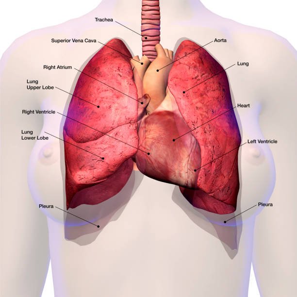
The primary organs of respiration are the lungs, or pulmones. The pleural sac encloses the right and left lungs, which are located in the thoracic cavity, one on each side of the mediastinum. The exchange of O2 and CO2 between inspired air and blood is the primary function of the lungs. With its base resting on the diaphragm and its peak extending into the root of the neck, each lung has a big conical or pyramidal form. Compared to the left lung, the right lung is heavier and larger. The left lung weighs 650 g and the right lung 700 g. The left lung possesses two lobes, while the right lung possesses three. Two lobar bronchi supply the lobes, which are divided by deep, noticeable fissures on the lung’s surface. The trachea and the heart are connected to the lungs by the main bronchi and pulmonary arteries, respectively.
EXTERNAL FEATURES :-
- Apex of the lungs
- Base of the lungs
- Border of the lungs
- Surface of the lungs
1. Apex of the lungs :-
The lung’s rounded or blunt superior end is its apex. It continues into the neck’s root around 2.5 cm above the medial third of the clavicle and 3 cm above the anterior end of the first rib. Each lung’s apex is covered by the visceral pleura, which joins the parietal pleura at the hilum. The pleural covering makes sure that there is no resistance against the thoracic cavity during respiration, allowing the lungs to expand and contract smoothly.
RELATIONS :- Anterior – (a). subclavian artery and vein (b). sternocleidomastoid artery (c). scalene muscle and Posterior -(a). first thoracic vertebra (b). ribs and intercostal muscle (c). sympathetic trunk (d). esophagus (e). trachea and main bronchi (f). brachial plexus
2. Base of the lungs :-
The base is also known as the diaphragmatic surface because it is the lower semilunar concave surface, which sits on the diaphragm’s dome. The diaphragm’s right dome divides the lung from the liver on the right side and its left dome divides the lung from the spleen and stomach fundus on the left side.
3. Borders of the lungs :-
The anterior, posterior, and inferior are the three primary borders of the lungs
(A). ANTERIOR:- The anterior border is the thin, pointed margin that extends along the front of the lung. The front boundary of the right lung extends from the apex to the level of the sixth costal cartilage. It approximates the sternum closely as it runs vertically along the midline. The cardiac notch is formed by the left lung’s front border, which also stretches from the apex to the sixth costal cartilage before veering laterally to pass around the fourth or fifth rib. The heart is accommodated by this notch, which results in a prominent depression.
(B). POSTERIOR :- The thick, rounded margin of the lung that runs down the rear is called the posterior border. It reaches from the C7 vertebral spine to the T10 vertebral spine. In imaging and physical examination, the link between the posterior border and the vertebral column is important, especially when assessing disorders such as lung tumors or pleural effusions.
(C). INFERIOR :- The lower edge of the lung that divides the base from the costal and mediastinal surfaces is known as the inferior border. It extends from the anterior to the posterior regions of the lung, following the diaphragm’s outline. At the sixth, eighth, and tenth ribs, respectively, the inferior border crosses the midclavicular, midaxillary, and scapular lines.
4. Surfaces of the lungs :-
Costal surfaces :- This is the part of the lungs’ exterior that faces the rib cage. It fits the contour of the ribs and is smooth and convex. It rests adjacent to the intercostal muscles and the ribcage on the inside of the thoracic wall. The lateral thoracic wall is involved. (In an embalmed and hardened lung, rib imprints can be seen on the costal surface.)
The following is the total number of ribs associated with this surface:
-The upper six ribs are located in the midclavicular line, the upper eight in the midaxillary line, and the top ten in the scapular line.
Diaphragmatic surface :- This surface, which sits on the diaphragm, is also referred to as the base of the lung. It conforms to the dome-shaped diaphragm by being concave. The liver is in contact with the diaphragmatic surface. The spleen and stomach are in contact with the diaphragmatic surface.
Medial surface :- It is separated into two sections: the massive anterior mediastinal portion and the little posterior vertebral half. Relations : The larger and lesser splanchnic nerves, posterior intercostal vessels, and the vertebral column are all connected to the vertebral portion. A hilum is seen in the mediastinal region, which is associated with mediastinal structures such the heart, large blood arteries, and nerves. Given that the mediastinum’s left and right surfaces have distinct structural compositions. Because the structures creating the right and left surfaces of the mediastinum differ, so do the relationships between the two lungs’ mediastinal surfaces. It is recommended that students become familiar with the structures that make up the left and right surfaces of the mediastinum in order to comprehend the relationships between the mediastinal surfaces of the lungs.
Structures that make up the mediastinum’s right surface :- 1. The right atrium makes up the majority of the right mediastinal surface.
2. The right brachiocephalic vein and superior vena cava are located above the right atrium.
3. The esophagus and trachea are located behind these structures.
4. The azygos vein is a sizable vein that ends in the superior vena cava after ascending along the side of the spinal column and arcing across the root of the right lung.
5. Three neurological structures: the right sympathetic chain, the right vagus nerve, and the right phrenic nerve. The phrenic nerve travels to the diaphragm, where it superficially contacts the right atrium, inferior vena cava, and superior vena cava, three venous systems that are located below. The lung’s root is in front of this passage. The right side of the trachea is where the vagus nerve crosses after passing behind the lung root. Here, it divides into branches that contribute to the development of the esophageal and posterior pulmonary plexuses. The paravertebral gutter is where the sympathetic trunk runs. The splanchnic nerves emerge from its bottom half, travel medially, and enter the belly via passing through the diaphragm’s crura.
Structures that make up the mediastinum’s left surface :- 1. The primary components that make up the left surface of the mediastinum are the aorta and left ventricle.
2. The aorta ascends initially, then drops behind the lung root after arching over the left lung root.
3. The brachiocephalic trunk, left subclavian vein, and left common carotid artery are the three main vessels that emerge from the aortic arch and proceed upward to the base of the neck.
4. The esophagus turns to the left behind the heart as it descends into the thorax, softly crossing the descending aorta’s path.
5. Three neurological structures: the left sympathetic chain, left vagus nerve, and left phrenic nerve -The diaphragm is reached by the left phrenic nerve after it crosses the aortic (left) side, goes in front of the lung root, and descends superficially into the left ventricle. l The aortic arch separates the left vagus nerve from the trachea. Here, it gives rise to the recurrent laryngeal branch, which ascends into the tracheoesophageal groove and hooks beneath the aortic arch. Runs behind the lung root below the aortic arch, the vagus nerve splits into the posterior pulmonary and esophageal branches. The sympathetic trunk and splanchnic nerves are positioned similarly to the right side.
Lobes and Fissures of the Lungs :-
The superior, middle, and inferior lobes of the right lung are separated by two fissures: an oblique fissure and a horizontal fissure. An oblique fissure separates the left lung into two lobes, superior and inferior.
Oblique fissure : A lengthy, oblique fissure crosses the inferior border approximately 7.5 cm (3 inches) lateral to the midline and the posterior border approximately 6 cm (2 inches) below the apex. It also travels obliquely downward and forward. It links the inferior lobe over the superior and middle lobes.
Horizontal fissure : Only one right lung has a brief horizontal fissure. It extends horizontally across to the anterior lung boundary from an oblique fissure at the midaxillary line. The middle and superior lobes are divided by it.
Root of the Lungs :-
The lung’s root is a wide, short pedicle that joins the mediastinum with the lung’s medial surface. It is made up of structures that enter and exit the lung at the hilum. The region located on the lung’s mediastinal surface via the structures that pass into or out of the lung is known as the hilum. A tubular sheath made of mediastinal pleura envelops the root of the lung. Root of the Right Lung positioned between the fifth and seventh thoracic vertebrae respectively.
Left Lung Root: Usually located between the fourth and sixth thoracic vertebrae, a little higher than the right.
Components : The following structures make up the lung’s root:
1. The left lung’s principal bronchus, and the right lung’s eparterial and hypoarterial bronchi.
2. Artery of the heart.
3. There are two pulmonary veins.
4. The bronchial arteries, two on the left and one on the right.
5. Veins in the bronchi.
6. The lung’s lymphatic system.
7. The nerves’ both anterior and posterior pulmonary plexuses.
INTERNAL FEATURES OF THE LUNGS :-
The primary components of the lung are the pulmonary units, which are involved in gas exchange inside the lung, and the intrapulmonary bronchial tree, which is involved in the conduction of air into and out of the lung.
Bronchial Tree :-
The complicated organize of aviation routes that supply the lungs with oxygen is called the bronchial tree. It starts in the trachea and partitions into lungs that house dynamically littler tubes. Principal bronchus, lobar bronchi, terminal bronchioles, and breathing bronchioles make up the bronchial tree.
TRACHEA :-
The windpipe, also referred to as the trachea, is an essential part of the respiratory system. It acts as the main conduit for speech to travel from the voice box, or larynx, to the bronchi, which then connects to the lungs. Beginning just below the larynx, the trachea grows until it reaches the fifth thoracic vertebra, at which point it splits into the right and removes the center bronchi. In adults, it measures approximately 10–12 cm (4-5 inches) in length and 2-2.5 cm (0.8–1 inch) in width. 16–20 C-shaped cartilaginous rings encircle the trachea, providing vital support and maintaining the opening of the flight path. The rings’ open portion faces the esophagus from the back. The ciliated pseudostratified columnar epithelium that lines the inside of the trachea is its lining. This coating hardens ciliated cells, which move actual fluid forward into the pharynx to be swallowed or ejected, and holder cells, which transfer substantial fluid to capture particles.
PRIMARY (MAIN) BRONCHUS :-
The carina, which is situated at the level between the fourth and fifth thoracic vertebra, is where the trachea divides into the left and right main bronchi.
Right primary bronchus – In comparison to the left bronchus, the primary right bronchus is larger, more vertical, and shorter (by about 2.5 cm). It splits immediately into three secondary bronchi (lobar bronchi), one for each of the superior, middle, and inferior lobes of the right lung.
Left primary bronchus – The left side’s primary bronchus is about 5 cm longer, narrower, and more horizontal.
It splits into two secondary bronchi (lobar bronchi), each of which serves the inferior and superior lobes of the left lung.
The left principle bronchus’ long axis deviates from the trachea’s long axis by approximately 45°, while the right principal bronchus’ long axis deviates from the latter by roughly 25°.
The left main bronchus descends beneath the aortic arch and crosses in front of the esophagus.
LOBAR (SECONDARY) BRONCHI :-
The right major bronchus splits (gives out) three lobar bronchi upon entering the lung, one for each of the right lung’s lobes. After entering the lung, the cleared out primary bronchus parts into two lobar bronchi, which are one for each cleared out lung projection.
TERTIARY (SEGMENTAL) BRONCHI :-
For every bronchopulmonary segment, the lobar bronchus splits into segmental (tertiary) bronchi. Repeated divisions of the segmental bronchi result in terminal bronchioles, which are extremely tiny bronchi. Respiratory bronchioles, which have walls devoid of cartilage, are produced by terminal bronchioles. A small area of the lung called a pulmonary unit, which is involved in gas exchange inside the lung, is ventilated by each respiratory bronchiole.
Smaller bronchioles split off from segmental bronchi. In contrast to bronchi, bronchioles are mostly made of smooth muscle and lack cartilage. They split into terminal bronchioles after that. The respiratory bronchioles are reached by these tiniest conducting airways. The respiratory zone of the lung, which is where gas exchange takes place, is indicated by these bronchioles. Alveolar ducts are where they lead. Alveolar ducts, which are encircled by alveoli, receive the output of the respiratory bronchioles. Gas exchange takes place in microscopic air sacs called alveoli. A network of capillaries encircles the incredibly thin alveolar walls.
ARTERIAL SUPPLY OF THE LUNGS :-
The two main arterial supply systems of the lungs are the pulmonary artery and the bronchial artery. Each system contributes differently to the overall well-being and maintenance of lung tissue.
Bronchial Artery :-
The bronchial tree and lung tissue receive nourishment from the bronchial arteries. One bronchial artery, which originates from either the upper left or right third posterior intercostal arteries, supplies the right lung. Two bronchial arteries, which emerge from the descending thoracic aorta, supply the left lung.
Pulmonary Artery :-
The lungs get deoxygenated blood from the pulmonary arteries. Every lung has its own pulmonary artery. These are the pulmonary trunk’s branches.
As they enter the hilum of their respective lungs, the right and left pulmonary arteries are located in front of the principal, or primary, bronchi. The left pulmonary artery sits inferior to the aortic arch at the level of the T5 vertebra, while the right pulmonary artery is crossed superior by the azygos vein arch. In the hilum, the pulmonary arteries split into lobar branches, which further split into segmental or terminal branches. The segmental branches grow in succession and match the bronchial tree’s segmental branches.
Venous Drainage :-
The pulmonary veins and the bronchial veins are the two systems that make up the venous drainage of the lungs.
Bronchial vein : The deoxygenated blood from the pulmonary tissue and bronchial tree is drained by the bronchial veins. Every side has two bronchial veins:
1. Azygos veins get drainage from the right bronchial veins.
2. The left superior intercostal vein or the hemiazygos vein receive the drainage from the left bronchial veins.
Pulmonary vein : The oxygenated blood in the lungs is expelled by the pulmonary veins. Every side has two pulmonary veins. Every vein in the body empties deoxygenated blood, with the exception of the pulmonary veins, which discharge oxygenated blood from the lungs.
• All of the body’s arteries carry oxygen, with the exception of the pulmonary arteries, which provide the lungs with blood that has lost oxygen and except for the pulmonary veins, which empty oxygenated blood from the lungs, each vein in the body drains deoxygenated blood.
• The bronchial arteries supply the bronchial tree up to the respiratory bronchioles, or non-respiratory sections of the lungs.
• The alveoli and pulmonary capillary beds feed the respiratory regions of the lungs with atmospheric air. The pulmonary arteries and veins do not travel together. While pulmonary artery branches are distributed segmentally, pulmonary vein tributaries are intersegmental in nature.
LYMPHATIC DRAINAGE OF THE LUNGS :-
Two sets of lymph arteries drain the lymph from the lung.
1. Superficial lymph vessels.
2. Deep lymph vessels.
1.Superficial lymph vessels : The peripheral lung tissue that is located underneath the visceral pleura is drained by these veins. Beneath the visceral pleura, they constitute the superficial (subpleural) plexus. The plexus vessels travel around the edges and boundaries of the lung fissures before arriving at the hilum, where they empty into the bronchopulmonary (hilar) lymph nodes.
2. Deep lymph vessels : These arteries create the deep plexus by drainage the blood vessels, connective tissue septa, and bronchial tree. The pulmonary arteries and bronchi from the deep plexus flow towards the lung’s hilum, passing through pulmonary lymph nodes found inside the lung material before draining into bronchopulmonary (hilar) lymph nodes.
Pathway : As a result, bronchopulmonary (hilar) lymph nodes receive drainage from both superficially and deep lymphatic plexuses. The superior and inferior tracheobronchial lymph nodes, which are situated superior and inferior to the tracheal bifurcation, respectively, receive lymph drainage from the hilar lymph nodes. These nodes eventually empty into the right lymphatic duct and the left thoracic duct on the right and left sides, respectively, as well as the pre- and paratracheal nodes of lymph and the rights and left bronchomediastinal lymphatic trunks.
NERVE SUPPLY OF THE LUNGS :-
Both of these nerve fibers supply the lung: Sympathetic and Parasympathetic
Parasympathetic fibers originate from the vagus nerve, whereas sympathetic fibers are produced by the spinal segments that correspond to T2 through T5. Both provide secretomotor function to the mucous glands of the respiratory tree and motor function to the breathing muscles. Vasodilatation, bronchoconstriction/bronchospasm, and increased mucous production are brought on by the parasympathetic fibers. Vasoconstriction, reduced mucous production, and bronchodilatation are all brought on by sympathetic fibers. Both sympathetic and parasympathetic fibers carry the afferent signal that originates from the bronchial mucous membrane and stretch receptors in the alveolar walls to the central nervous system.