
Cell Junctions
Introduction :-
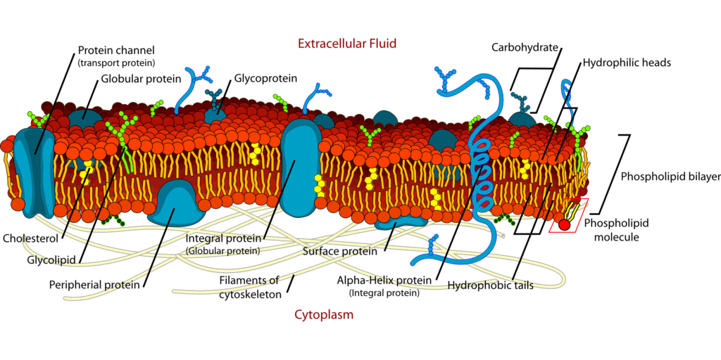
Cell Junctions- A cell connection is a connection between adjacent cells or a contact between a cell and the extracellular matrix. They play important roles in maintaining the structural integrity of tissues, facilitating communication between cells, and controlling the passage of substances. Also called a membrane connection.
Cell connections are classified into three types:
1. Occlusion connections,
2. Communication connections, and
3. Anchoring connections.
1. Cell Junctions- Occlusion connections :-
Cell connections that prevent the intercellular exchange of substances are called occlusive connections. These compounds prevent ions and molecules from moving between cells. Ex. Tight connections
Cell Junctions- Tight Junction :-
Large molecules cannot flow through tight junctions because they are occlusive connections.They are also called zonula occludens. They are areas where the cell membranes of adjacent cells are tightly fused. This type of connection is present in the apical ends of epithelial and endothelial cells of the intestinal mucosa, in the walls of renal tubules, in the capillary walls and in the choroid plexus.
Structure of Tight Junction :-
A tight junction consists of a ridge with two halves: one half of the ridge comes from one cell and the other half comes from the other cell. Both halves of the ridge join very tightly together and occupy the space between the two cells. Each half of the ridge is made up of tight junction strands.
Protein Involve Tight Junction :
1. Claudins: These are the main structural proteins of tight junctions. They form strands that seal the space between cells and determine the permeability properties of the connection.
2. Occludins: These proteins interact with claudins and contribute to the stability and integrity of tight junctions.
3. Junctional adhesion molecules (JAMs): These proteins are involved in cell-cell adhesion and help form and maintain tight junctions.
4. Zonula occludens (ZO) proteins: These intracellular scaffolding proteins (ZO-1, ZO-2, and ZO-3) connect the transmembrane proteins of tight junctions to the actin cytoskeleton, providing structural support.
Functions of Tight Junction :-
1. Stability and strength: Tight connections provide tissues with stability and strength by tightly holding nearby cells together.
2. Selective permeability (gating function): tight connections form a selective barrier to small molecules and a complete barrier to larger molecules. In epithelial and endothelial cells, tight junctions are the best intercellular junctions, and act as a selective (semi-permeable) diffusion barrier between adjacent cells. This function is called the barrier or gate function. The barrier function of tight junctions regulates the exchange of ions, water, and various macromolecules between cells. Each tissue has this role to a different degree.. In some epithelial cells, only a few substances pass through the tight junctions (by diffusion or active transport). In other cells, no substances pass through the tight junctions.
3. Fence function: Tight junctions prevent the lateral movement of proteins (integral membrane proteins) and lipids within the plasma membrane, thus acting as a fence. The fence function maintains the distinct composition of proteins and lipids between the apical and basolateral plasma membrane domains. Because of this feature, tight junctions are sometimes called impermeable junctions.
4. Preservation of cell polarity: Proteins are held in the apical portion of the cell membrane by the fencing function of tight junctions.
5. Blood-brain barrier: The blood-brain barrier is formed by tight connections in brain capillaries and keeps a variety of chemicals from penetrating brain tissue from capillary blood. Only lipid-soluble substances medicines and steroid hormones, for example, are able to pass past the blood-brain barrier.
Applied Physiology of Tight Junction :-
Diseases caused by mutations in genes that code for tight junction proteins:
1. Hereditary deafness
2. Ichthyosis (scaly skin)
3. Sclerosing cholangitis (constipation-related bile duct inflammation)
4. Hereditary hypomagnesemia (low magnesium levels in the blood)
5. Synovial sarcoma (soft tissue cancer) Tight junction function is also affected by some bacteria and viruses.
2. Cell Junctions- Communication connections :-
Cell connections that allow for the cell-cell exchange of substances are called communication connections i.e. These compounds allow ions and molecules to move between cells. Gap junctions and chemical synapses are communication connections.
1. Cell Junctions- Gap junctions :-
Connections between cells known as gap junctions, or nexuses, permit the movement of ions and other tiny molecules between them. They are found in the basal layers of epithelial cells in the heart and in the intestinal mucosa.
Structure of Gap Junction:-
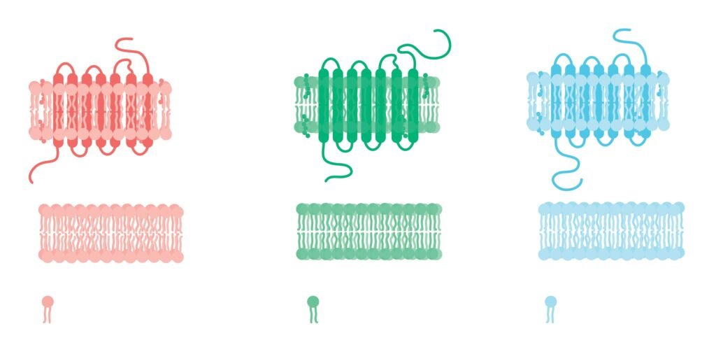
In vertebrates, gap junctions are formed by proteins called connexins (innexins in invertebrates). Six connexins together form a hemichannel or connexon in the cell membrane that aligns with a connexon in an adjacent cell to form a complete gap junction channel.
Function of Gap Junction:-
1. The diameter of the gap junction channel is about 1.5-3 nm, so the channel allows the passage of substances with less than molecular weight like glucose, amino acids, ions etc.
2. It helps in the exchange of chemical messengers between cells.
3. It helps in the rapid spread of action potentials from one cell to another.
2. Cell Junctions- Chemical Synapses :-
A chemical synapse is a kind of synapse where neurotransmitters are released to allow communication between neurons (or with other types of cells, including muscle cells). These chemicals, known as neurotransmitters, act as messengers between neurons, bridging a narrow opening known as the synaptic cleft to transmit messages.
Structure of Chemical Synape :-
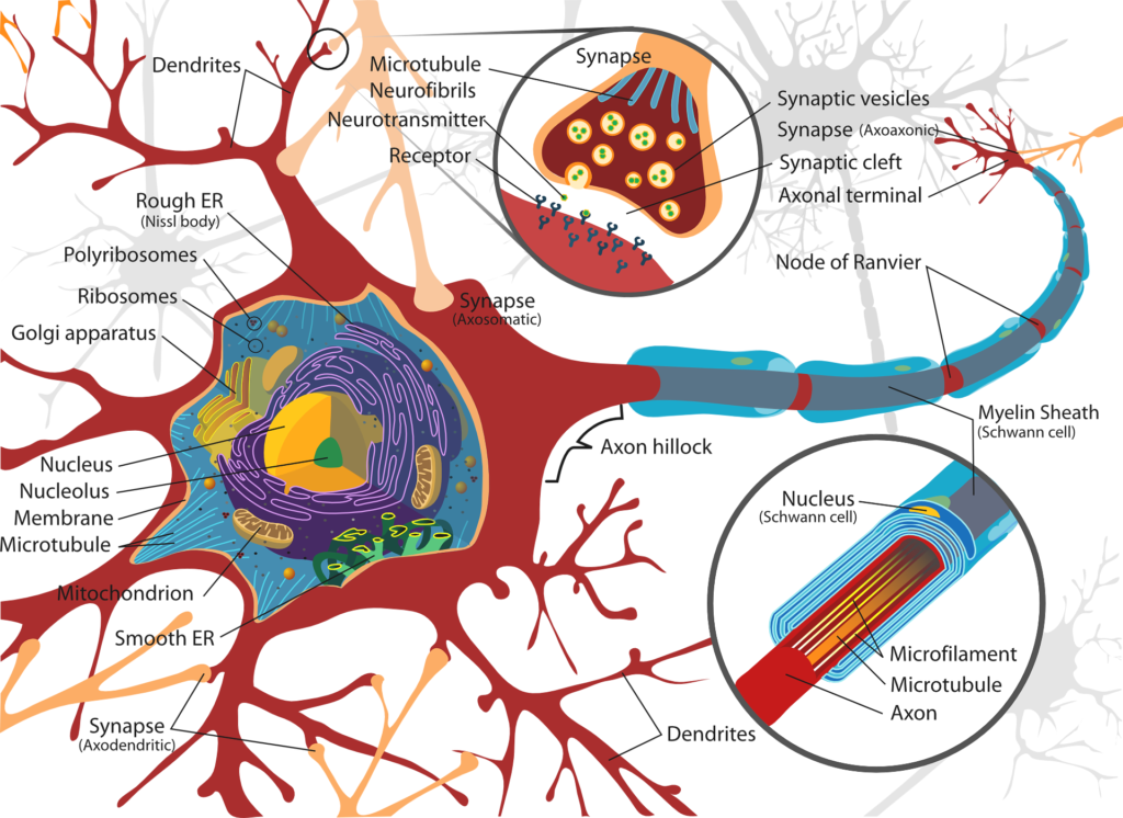
A typical chemical synapse contains the following main components:
• Presynaptic neuron: The signal-sending neuron. Neurotransmitter-filled synaptic vesicles are present.
• The narrow space separating presynaptic and postsynaptic neurons is known as the synaptic cleft. It is usually about 20-40 nanometers wide.
• Postsynaptic neuron: The neuron (or muscle or gland cell) that receives the signal. Its membrane has receptors that bind to neurotransmitters released by the presynaptic neuron.
3. Cell Junctions- Anchoring connections :-
These are desmosomes (which connect the cell’s intermediate filaments to other cells), hemidesmosomes, and anchor connections. Desmosomes, also known as macular adherens, are specialized cellular structures for cell-cell adhesion. The cell adhesion proteins in desmosomes are members of the cadherin family. Hemidesmosomes look like half-desmosomes that connect cells to the basement membrane below. Hemidesmosomes use the Desmopenetrin is a member of the integrin family of cell adhesion proteins. The adhesive compound has an anchoring function through its cytoplasmic actin filaments.
Applied Physiology :-
1. Dysfunction of colon adhesions and focal junctions due to protein mutations causes colon cancer and also tumor metastasis (spread of cancer cells from the primary tumor to other parts of the body)
2. Desmosome dysfunction causes bullous pemphigoid, an autoimmune disease characterized by a skin rash with raised blisters. Patients with this disease produce antibodies against cadherins
3. Hemidesmosome dysfunction can also cause bullous pemphigoid, as patients develop antibodies against integrins.